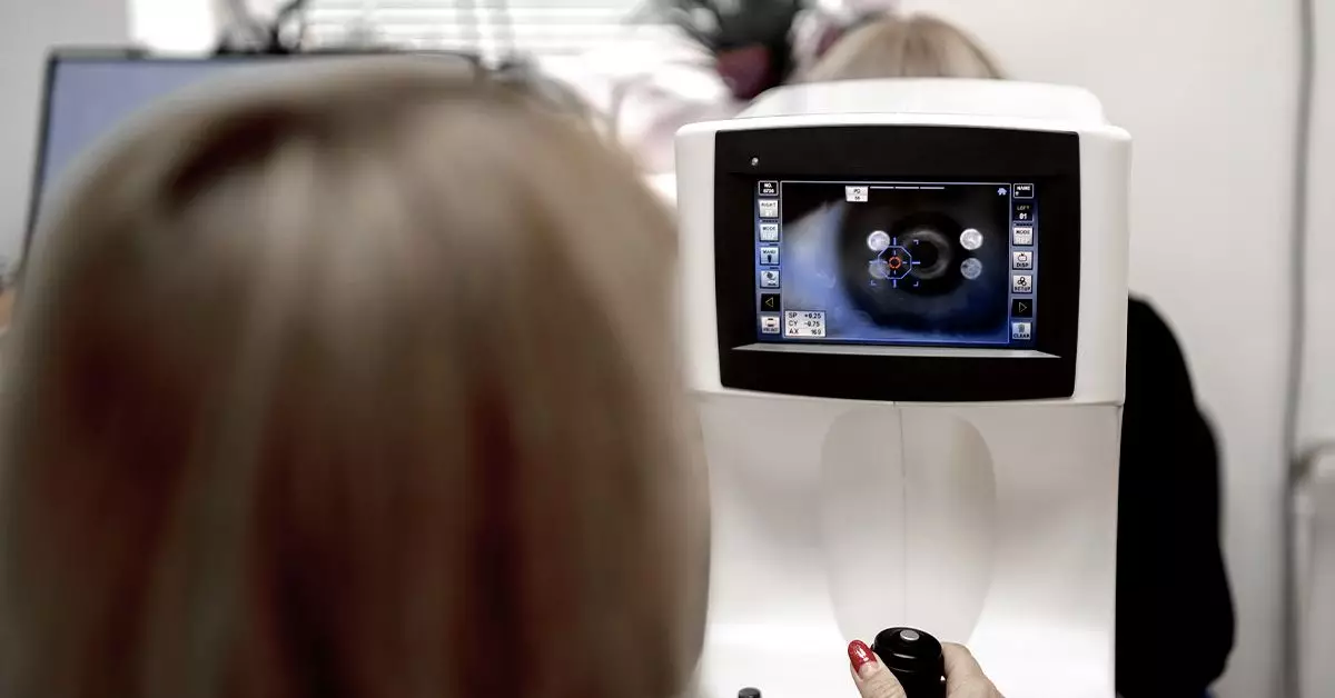
Diabetic retinopathy remains an alarming concern for many individuals living with diabetes, as it poses a significant risk of vision loss. A crucial tool in combating this condition is fundus photography, a technique that captures detailed images of the interior of the eye. This article delves into the intricacies of fundus photography, its relevance in detecting diabetic retinopathy, and the importance of timely eye examinations.
Fundus photography serves as a critical component of an examination known as fundoscopy, which scrutinizes the back of the eye, or fundus. This imaging captures detailed views of the retina, offering a glimpse into the health of the eye’s internal structures. Through the use of specialized cameras, healthcare professionals can visualize any potential abnormalities that may signify the onset of diseases like diabetic retinopathy.
Diabetic retinopathy arises from fluctuations in blood sugar levels over time, affecting the retina’s blood vessels. It can progress silently, often without noticeable symptoms until significant damage has occurred. Therefore, employing fundus photography for early diagnosis is vital for preventing irreversible vision damage.
When evaluating fundus photographs for signs of diabetic retinopathy, healthcare providers look for specific indicators:
1. **Microaneurysms**: These minuscule bulges in the retinal blood vessels manifest as small red dots and signify the initial stage of diabetic retinopathy. They are crucial for diagnosis, especially in patients with Type 1 and Type 2 diabetes.
2. **Hemorrhages**: Damage to the fragile blood vessels can lead to bleeding within the retina. Such hemorrhages may appear in different shapes—flame-shaped or dot-and-blot—and can notify the healthcare provider of the disease’s severity.
3. **Exudates**: Hard exudates, appearing as yellow or white patches, result from lipid deposits leaking from damaged blood vessels. Their presence can indicate swelling in the retina or ongoing blood vessel changes.
4. **Cotton Wool Spots**: These fluffy white patches signal areas of retinal nerve fiber swelling, often due to inadequate blood supply. Observing such spots can help track the disease’s progression.
5. **Neovascularization**: The abnormal growth of new blood vessels signifies more advanced diabetic retinopathy. This stage can lead to severe complications, including vision loss.
Alongside these visible indicators, there are subtler changes such as intraretinal microvascular abnormalities (IRMA), which highlight the irregularity in retinal blood vessels.
Recent advancements have transformed how we approach eye screening, notably through Selfie Fundus Imaging (SFI). This innovation empowers individuals to utilize their smartphones to capture images of their retina after applying dilating eye drops. A 2022 study underscored the effectiveness of this method by comparing three techniques of fundus photography. Remarkably, images taken by patients, following a brief instructional video, were comparable to those taken by trained professionals.
SFI presents a promising alternative, expanding the accessibility of diabetes monitoring and possibly reducing healthcare burdens. With the convenience of smartphones, proactive retinal health maintenance could become more commonplace.
Despite the technological advances in fundus photography, one critical factor remains: the need for regular eye exams. Adults with diabetes should schedule comprehensive dilated eye exams at least once a year. Early detection through routine screenings can drastically reduce the risks associated with diabetic retinopathy—up to 90% of vision loss from the disease may be preventable with timely intervention.
For those who may become pregnant or develop gestational diabetes, immediate eye assessments are indispensable. Pregnancy can lead to fluctuating blood sugar levels, increasing the likelihood of rapid progression in retinal disease.
Many individuals may not experience any symptoms, particularly in the early stages of diabetic retinopathy. However, changes like fluctuating vision, floaters, or a persistent feeling of blurred sight warrant urgent consultation with an eye care professional. By maintaining awareness of potential visual disturbances, patients can act swiftly to seek medical assistance and minimize the risk of complications.
While fundus photography plays a pivotal role in early detection and management of diabetic retinopathy, it is paramount to combine it with overall diabetes management. Regular physical activity, a balanced diet, and adherence to prescribed medications are all part of an effective strategy that significantly mitigates the risk of eye-related complications.
Fundus photography encompasses a powerful tool in the fight against diabetic retinopathy. Its capacity to reveal critical signs of eye disease underscores the importance of regular eye examinations. As technology evolves, the incorporation of methods like Selfie Fundus Imaging extends the possibilities for proactive health management. Individuals with diabetes must embrace these advancements and prioritize their retinal health to preserve their vision.

