
In the realm of diagnostic testing, particularly for breast cancer, the terms sensitivity and specificity serve as the yardsticks by which the effectiveness of screening methods are measured. A systematic review published in 2019 highlighted that ultrasound boasts an impressive sensitivity rate of 80.1% and a specificity of 88.4%. Sensitivity, in layman’s terms, is the tool’s ability to correctly flag those who actually have the disease, ensuring that few cases slip through the cracks. Conversely, specificity reflects how deftly a test can identify individuals who do not have the condition, avoiding unnecessary anxiety and follow-up procedures. When combined, these metrics speak volumes about an ultrasound’s reliability in the early detection of breast cancer, and it is vital for health practitioners to leverage this robust data in patient care.
Situations that Call for Ultrasound Screening
Ultrasound imaging has its own distinct niche in breast cancer detection protocols. According to Cancer Research UK, the application of breast ultrasounds is recommended in various scenarios: when a lump presents itself but goes undetected on a mammogram, to clarify whether a lump is solid or fluid-filled, and even as a regular screening tool for those with dense breast tissue lacking apparent lumps. This versatility makes ultrasound an indispensable adjunctive tool alongside the mammogram—particularly when the latter fails to produce clear results. In fact, the integration of both tests can significantly enhance diagnostic accuracy and facilitate timely interventions for individuals who may be at risk.
Step-by-Step: What to Expect During an Ultrasound
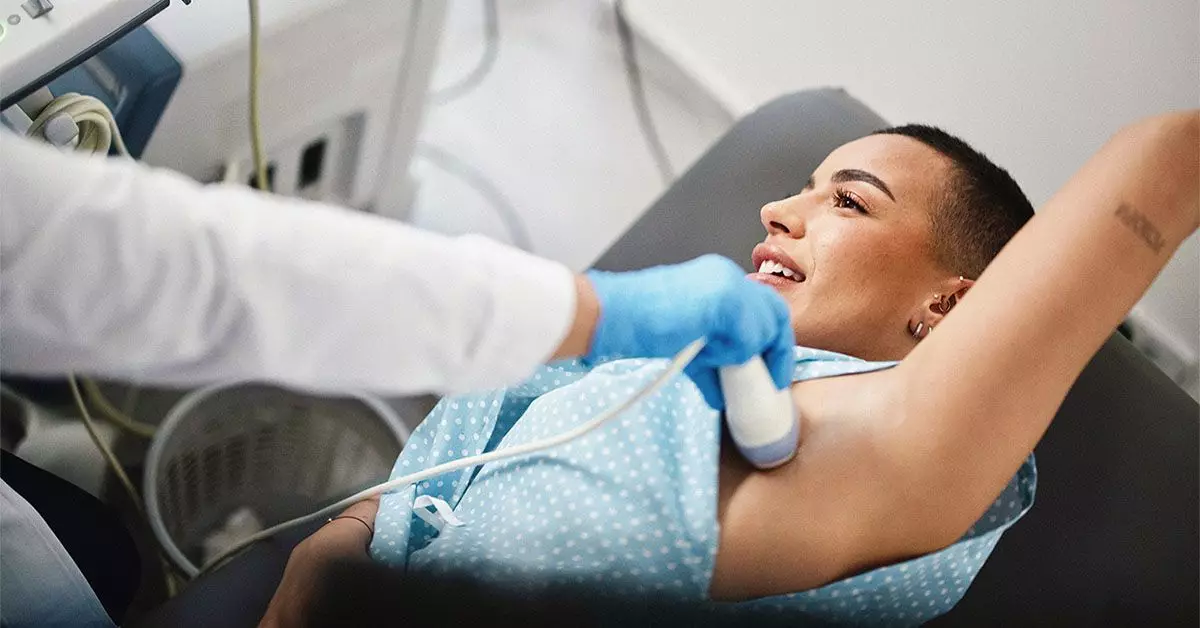
For those scheduled for a breast ultrasound, an understanding of the procedure can help alleviate apprehension. The process begins with the patient removing their clothing from the waist up, allowing the sonographer access to the area of interest. The application of a lubricating gel is crucial; it ensures that the ultrasound probe glides smoothly over the skin, generating high-quality images. The sonographer meticulously maneuvers the probe across the breast and into the armpit region to examine lymph nodes, which can often hold crucial information regarding cancerous spread. Such thoroughness in imaging not only aids in diagnosis but also informs future treatment decisions.
The Importance of Proactive Screening
Healthcare professionals consistently urge individuals to pay attention to bodily changes and make proactive decisions regarding screenings. The American Cancer Society recommends that individuals at average risk for breast cancer begin annual screenings at the age of 45, while those between 40 and 44 should consider initiating yearly mammograms. However, the presence of alarming symptoms, such as lumps, necessitates immediate consultation with a healthcare provider. Early detection through these screenings dramatically increases survival chances and treatment efficacy, highlighting the essential role of informed health choices in breast cancer management.
As technology advances, tools such as ultrasounds become pivotal in the fight against breast cancer. With high sensitivity and specificity rates and varied applications in diagnosis and monitoring, this imaging technique empowers both patients and clinicians to engage in more informed healthcare choices.