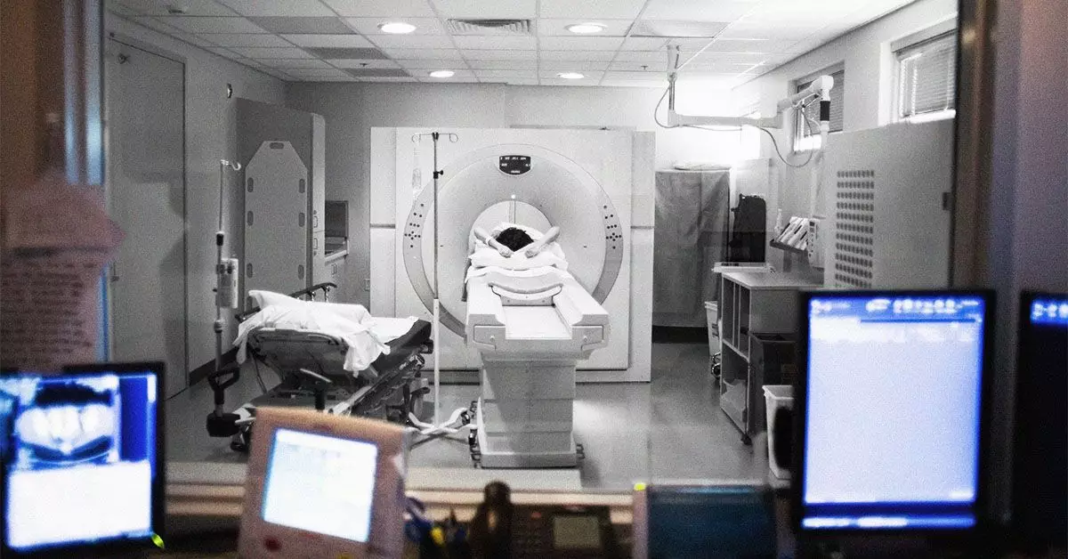
Computed Tomography (CT) scans are invaluable imaging tools that have transformed modern medicine’s approach to diagnostics. These scans employ a sophisticated technique that utilizes X-rays to produce detailed, cross-sectional images of the human body. While often employed to identify injuries or various diseases, their application in the realm of breast cancer diagnosis is limited and not a standard practice among healthcare providers. Nevertheless, certain scenarios may lead to incidental findings that could uncover signs of breast cancer, warranting closer examination.
CT scans generate a series of two-dimensional images that physicians can either view individually or utilize to construct a three-dimensional representation of the area in question. This detailed imagery allows medical professionals to discern irregularities pertaining to bones, organs, and soft tissues, facilitating a more comprehensive diagnostic process. Typically, a contrast dye injects into the patient’s bloodstream, enhancing the clarity of the imagery, particularly for soft tissues and vascular structures. Depending on the context, these contrast agents might be delivered intravenously or through oral intake, such as barium solutions.
Despite not being the primary diagnostic tool for breast cancer, studies indicate that CT scans can reveal incidental findings that might suggest the presence of breast malignancies. A noteworthy 2021 study illustrated that as many as 28% of CT scans may inadvertently identify cancerous breast lesions. This incidental detection can pose both a boon and a bane for patients. While it can lead to early diagnosis, the reliance on incidental findings raises questions regarding the efficacy of relying on CT imaging as a reliable standard for breast cancer diagnostics.
Interestingly, non-contrast CT scans may sometimes outperform mammography in identifying breast lesions, especially in cases where dense breast tissue obscures issues. The challenge lies in the fact that while CT scans may reveal lesions appearing to be cancerous, they should not be entirely substituted for more traditional and systematic diagnostic practices, such as mammograms.
Screening and Diagnostic Alternatives
Mammography remains the gold standard for breast cancer screening. This method uses low-dose X-rays to illuminate potentially problematic areas within breast tissue while minimizing radiation exposure. For individuals presenting with symptoms or abnormalities discovered during screening, diagnostic mammograms offer a more nuanced examination, providing additional clarity concerning the condition of the breast tissue.
Complementary imaging modalities exist, each equipped with unique strengths. Breast ultrasounds utilize sound waves to generate images and are particularly beneficial for assessing dense breast tissue. Magnetic Resonance Imaging (MRI) is another effective method that employs magnets and radio waves for detailed imaging, enhancing the physician’s understanding of the tumor characteristics.
Moreover, biopsies play a critical role in confirming breast cancer diagnoses. By extracting tissue samples from suspicious areas, healthcare providers can analyze the cellular structure at a microscopic level, leading to definitive diagnoses.
Symptoms and Screening Importance
Awareness of breast cancer symptoms is paramount. Changes such as new lumps, mass formations, or alterations in the breast or nipple should beckon the attention of healthcare professionals. Although some symptoms may indicate benign conditions, seeking medical advice promptly is essential for identifying the underlying causes and potentially implementing necessary treatment.
Routine breast cancer screenings represent a fundamental component of preventive healthcare, often detecting abnormalities before symptoms emerge. Early intervention is linked to significantly more favorable outcomes, underscoring the importance of attending scheduled screenings and consultations with medical professionals.
Despite advancements in imaging technology, a 2023 study reported concerning statistics regarding the ability of radiologists to detect incidental breast cancer findings on routine chest CT scans. Astonishingly, the study revealed that 64.3% of breast cancer cases were overlooked during these assessments, particularly smaller lesions that were less conspicuous. Given that most chest CT scans inevitably display some form of breast tissue, the ramifications of missed findings can significantly impact patient health outcomes.
Indications of potential breast cancer observable on CT scans may manifest as irregularly shaped masses exhibiting surface spikes or exhibiting rim enhancement when contrast dye is applied. Such characteristics necessitate meticulous interpretation, reinforcing the argument that while CT scans may occasionally unveil troubling signs, mammograms and other imaging modalities should remain at the forefront of breast cancer screening.
While CT scans can yield incidental insights into breast cancer, they should reinforce rather than replace traditional diagnostic approaches. The incorporation of mammography, ultrasound, and MRI provides a comprehensive strategy for evaluating breast health. As research continues to evolve, medical professionals are increasingly urged to examine CT scans with a keen eye for incidental findings that could indicate malignancy. Prioritizing communication with healthcare providers after observing unusual breast changes is vital, ensuring early intervention and improved prognostic outcomes.

